Research
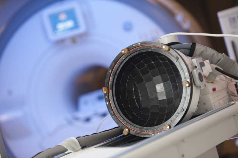
During my PhD, I worked with focused ultrasound, a technology used to perform non-invasive brain surgery in humans. I've also performed research in neurosurgery and nuclear medicine.
Improving noninvasive brain surgery
Noninvasive brain surgery using focused ultrasound is already being performed in hospitals, though targeting structures inside the brain can still be improved. Using the rapid beam simulation framework I previously developed, I showed that the technique provides great promise for improving treatment outcomes
A rapid beam simulation framework
Focused ultrasound is used in noninvasive brain surgery to heat up and destroy brain tissue. Before doing irreversible damage, the surgeon would like to know where the focal spot is. I've developed a simulation framework that predicts where the focal spot will be and how hot it will be, with the potential to be used in real-time during treatments
Understanding complications of laser thermal therapy
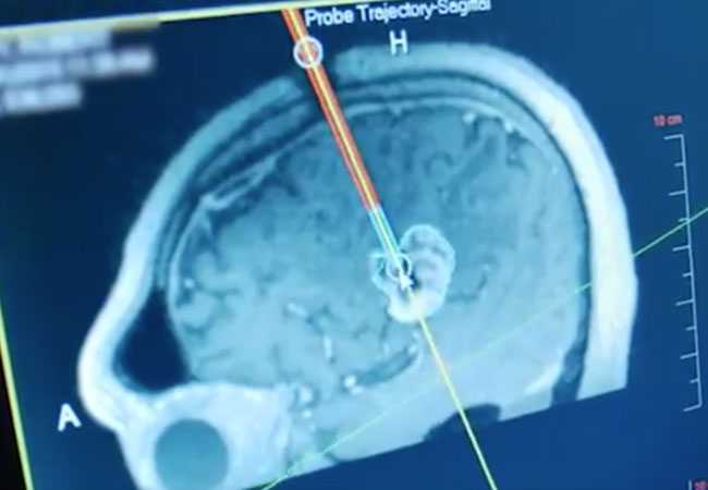
Laser interstitial thermal therapy is a minimally invasive therapy for treating medication-resistant epilepsy. There have been several cases of adverse complications due to this therapy, and in this work we investigated the likely causes of these complications
Individualized estimates of radiation exposure
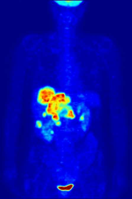
Patients who undergo positron emission tomography imaging are exposed to ionizing radiation. However, patients' exposure to radiation is not well quantified. In a push towards personalized medicine, we performed estimations of radiation exposure on a patient-specific basis
Understanding attenuation in skull bone
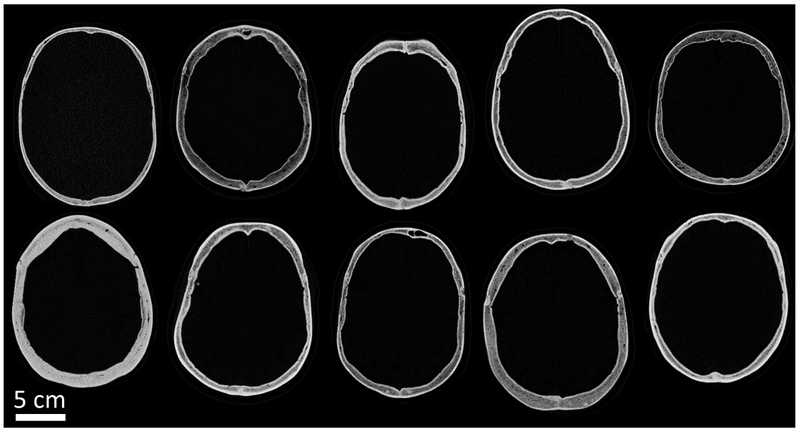
Accurate simulations of ultrasound propagation depend on accurate simulation parameters. The skull is a highly complex medium that distorts the ultrasound focal spot and displaces it away from the target. We performed empirical measurements of attenuation in human skull bone in order to improve simulation accuracy
Examining alternative methods for treatment planning
Treatment planning has traditionally been performed using computed tomography (CT) images, though magnetic resonance (MR) images are an attractive alternative. In this study, we investigated whether MR can perform as well as the gold standard CT
Understanding acoustic velocity in skull bone
Accurate simulations of ultrasound propagation depend on accurate simulation parameters. The skull is a highly complex medium that distorts the ultrasound focal spot and displaces it away from the target. We performed empirical measurements of acoustic velocity in human skull bone in order to improve simulation accuracy
Extracting additional information from skull bone
Although computed tomography (CT) images have traditionally been used for treatment planning, magnetic resonance (MR) images are an attractive alternative. This study examined the capacity of MR to supplement or replace CT as a means of estimating acoustic velocity of the skull

